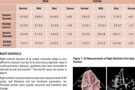
normal lv size | normal Lv dimensions normal lv size Each echocardiogram includes an evaluation of the LV dimensions, wall thicknesses and function. Good measurements are essential and may have implications for therapy. The LV dimensions must be measured when the end-diastolic and end-systolic valves (MV and AoV) are closed in the parasternal long axis (PLAX) view. LV 452 Euro IV. BUS LV 452. El bus LV 452, está diseñado para servicio intermunicipal y especial y da cumplimiento a las nuevas reglamentaciones de accesibilidad y seguridad.
0 · normal Lv size and function
1 · normal Lv end diastolic diameter
2 · normal Lv dimensions
3 · normal Lv diameter
4 · left ventricular wall thickness chart
5 · left ventricular diameter chart
6 · left ventricle size chart
7 · Lv wall thickness normal values
Chris Rolland, CEO. Chris joined AllClear in January 2018 as CEO, with over 20 years Financial Services experience. Previously he was CEO of Staysure and American Express Insurance Services. He holds an MBA, BA Economics and FS qualifications from the Chartered Institute of Insurance, and the Institute of Financial Services.
Each echocardiogram includes an evaluation of the LV dimensions, wall thicknesses and function. Good measurements are essential and may have implications for .Normal Ranges for LV Size and Function Normal values for LV chamber dimensions (linear), volumes and ejection fraction vary by gender. A normal ejection fraction is 53-73% (52-72% for men, 54-74% for women). Refer to Table 2 (normal values for non-contrast images) and Table 4 (recommendations for the normal
Each echocardiogram includes an evaluation of the LV dimensions, wall thicknesses and function. Good measurements are essential and may have implications for therapy. The LV dimensions must be measured when the end-diastolic and end-systolic valves (MV and AoV) are closed in the parasternal long axis (PLAX) view. ∗ LV size applied only to chronic lesions. Normal 2D measurements: LV minor axis ≤ 2.8 cm/m 2 , LV end-diastolic volume ≤ 82 ml/m 2 , maximal LA antero-posterior diameter ≤ 2.8 cm/m 2 , maximal LA volume ≤ 36 ml/m 2 (2;33;35).Taking the example of LV dimensions: just as 2.3% of normal individuals will have an LV size above the upper reference limit, a tiny proportion of normal individuals (just 0.15% of the normal population) would have values in the ‘moderate’ range using this methodology. The majority of these studies were focused on left ventricular (LV) cavity size, 3 – 5, 7 –10,12 reference values for atrial and right ventricular dimensions barely exist. 6, 11. Table 1. Publications on Normal Values of Left Ventricular Cavity Dimensions by Echocardiography.
Classifying LV size. Defining normal values for ventricular size is important for the standardisation of echocardiographic reporting, but is not a straightforward task. Normal and abnormal ranges depend on a number of factors including populations studied, methods used for imaging and the statistical approaches employed.
LV size was categorized by using either LV end-diastolic or end-systolic diameter or a qualitative assessment, as follows: normal, smaller than 4 cm; mildly enlarged, 4.1 to 5.4 cm moderately enlarged, 5.5 to 6.5 cm; and severely enlarged, larger than 6.5 cm.
Normal values of left ventricular mass (LV M) and cardiac chamber sizes are prerequisites for the diagnosis of individuals with heart disease. LV M and cardiac chamber sizes may be recorded during cardiac computed tomography angiography (CCTA), and thus modality specific normal values are needed. Uncontrolled high blood pressure is the most common cause of left ventricular hypertrophy. Complications include irregular heart rhythms, called arrhythmias, and heart failure. Treatment of left ventricular hypertrophy depends on the cause. Treatment may include medications or surgery. Mean normal values for indexed end-diastolic volume, end-systolic volume, and LVEF in men and women were 70 ± 15 and 65 ± 12 mL/m 2, 28 ± 7 and 25 ± 6 mL/m 2, and 60 ± 5% and 62 ± 5%, respectively. Men had larger LV volumes and lower LVEFs than women.
Normal Ranges for LV Size and Function Normal values for LV chamber dimensions (linear), volumes and ejection fraction vary by gender. A normal ejection fraction is 53-73% (52-72% for men, 54-74% for women). Refer to Table 2 (normal values for non-contrast images) and Table 4 (recommendations for the normal Each echocardiogram includes an evaluation of the LV dimensions, wall thicknesses and function. Good measurements are essential and may have implications for therapy. The LV dimensions must be measured when the end-diastolic and end-systolic valves (MV and AoV) are closed in the parasternal long axis (PLAX) view.
∗ LV size applied only to chronic lesions. Normal 2D measurements: LV minor axis ≤ 2.8 cm/m 2 , LV end-diastolic volume ≤ 82 ml/m 2 , maximal LA antero-posterior diameter ≤ 2.8 cm/m 2 , maximal LA volume ≤ 36 ml/m 2 (2;33;35).
Taking the example of LV dimensions: just as 2.3% of normal individuals will have an LV size above the upper reference limit, a tiny proportion of normal individuals (just 0.15% of the normal population) would have values in the ‘moderate’ range using this methodology. The majority of these studies were focused on left ventricular (LV) cavity size, 3 – 5, 7 –10,12 reference values for atrial and right ventricular dimensions barely exist. 6, 11. Table 1. Publications on Normal Values of Left Ventricular Cavity Dimensions by Echocardiography.
Classifying LV size. Defining normal values for ventricular size is important for the standardisation of echocardiographic reporting, but is not a straightforward task. Normal and abnormal ranges depend on a number of factors including populations studied, methods used for imaging and the statistical approaches employed.LV size was categorized by using either LV end-diastolic or end-systolic diameter or a qualitative assessment, as follows: normal, smaller than 4 cm; mildly enlarged, 4.1 to 5.4 cm moderately enlarged, 5.5 to 6.5 cm; and severely enlarged, larger than 6.5 cm.
Normal values of left ventricular mass (LV M) and cardiac chamber sizes are prerequisites for the diagnosis of individuals with heart disease. LV M and cardiac chamber sizes may be recorded during cardiac computed tomography angiography (CCTA), and thus modality specific normal values are needed. Uncontrolled high blood pressure is the most common cause of left ventricular hypertrophy. Complications include irregular heart rhythms, called arrhythmias, and heart failure. Treatment of left ventricular hypertrophy depends on the cause. Treatment may include medications or surgery.
normal Lv size and function

michael kors vanilla belt
michael kors táska fehér

This synthetic foam concentrate is intended for forceful or gentle firefighting applications at 3% solution on hydrocarbon fuels and at 3% solution on polar solvent fuels in fresh, salt, or hard water. CHEMGUARD C334-LV foam solution utilizes three suppression mechanisms intended for rapid fire knockdown and superior burnback resistance:
normal lv size|normal Lv dimensions


